A harmless wave of sound with no radiation will reveal what is underneath, saving probably many lives. This is the power of breast ultrasound-an essential tool in our fight against breast cancer.
Mammograms are the best way to check for breast cancer, although ultrasound technology is becoming a very useful additional tool. It has been particularly true of women who have dense breast tissue, or sometimes people need extra information. It’s a safe and non-invasive method that has altered the ways by which we find, diagnose, and keep track of breast problems. It gives live images that can really help in finding issues early.
Understanding the tools and techniques for detecting breast cancer is vital for everyone. To learn more about breast cancer causes, symptoms, and treatment options, visit our comprehensive guide here. Staying informed is your first step towards better breast health.”
Let’s look at how ultrasound technology is changing the way we detect and treat breast cancer. We will explain the basic ideas and the newest advancements. We will tell you everything you need to know about breast ultrasound, including its benefits, uses, limitations, and what to expect during the procedure.

Understanding Breast Ultrasound Basics
How Breast Ultrasound Functions
Breast ultrasound uses high-frequency sound waves to make clear images of breast tissue. A handheld tool called a transducer sends out these waves, which bounce off breast parts and come back to make live images on a screen. This method does not involve surgery and helps radiologists tell the difference between fluid-filled cysts and solid lumps.
Differences from Mammography
Key differences between ultrasound and mammography include:
- Ultrasound does not use radiation
- Improved way to see dense breast tissue
- Real-time imaging capability
- More Comfortable procedure without compression
- Portable and flexible scanning angles
| Feature | Ultrasound | Mammography |
| Radiation | None | Uses X-rays |
| Best for | Dense breasts, younger women | Calcifications, overall screening |
| Image type | Real-time, dynamic | Static 2D or 3D |
| Compression | Not required | Required |
Types of Ultrasound Technology Used
The modern-day breast ultrasound systems have:
Automated Whole Breast Ultrasound (ABUS)
- Standardized imaging
- Complete coverage
- Reduced operator dependency
Handheld Ultrasound
- Traditional method
- Targeted examination
- Immediate help for biopsies
Doppler Ultrasound
- Blood circulation visualization
- Tumor vascularity determination
Having understood all these concepts, let’s try to explore the benefits offered by these technologies in breast cancer detection. Additionally, for men concerned about breast health, understanding male breast cancer is equally vital. Learn more about its causes, symptoms, and treatment options in our complete guide. Early awareness saves lives.
Major Advantages of Ultrasound in Breast Cancer Detection
Finding Problems Early
- Detects as small as 2-3 millimeters
- Particularly effective for dense breast tissue
- Can distinguish between solid masses and fluid-filled cysts
- It helps identify cancers that mammograms might miss
Non-invasive and Radiation-free Procedure
- No radiation exposure, unlike mammograms.
- Safe for pregnant women and regular check-ups
- Painless examination without compression
- No recovery time needed
Real-time Imaging Advantages
- Immediate visualization of breast tissue
- Active assessment of how blood flows
- Allows precise needle guidance during biopsies
- Interactive testing with rapid responses
Cost-Effectiveness in Comparison with Other Methods
| Screening Method | Average Cost | Time Required | Radiation Exposure |
| Ultrasound | $100-300 | 15-30 mins | None |
| Mammogram | $200-400 | 30-45 mins | Yes |
| MRI | $1000-2000 | 45-60 mins | None |
Breast ultrasound is an excellent finding and diagnostic tool in early-onset breast cancers. It can produce clear images of the breast tissue without causing any damage to them in real time. It’s just perfect for regular check-ups as well as for diagnosis. Since no radiation is exposed, it is harmless to young women and pregnant patients as well as to people who need to be checked frequently. Additionally, its relatively low cost as compared to other imaging methods makes very important breast cancer screening services reach more people. The above benefits, along with being very compatible with other imaging methods, make ultrasound a very integral part of comprehensive breast cancer detection plans.
To better understand how breast ultrasound integrates with other screening methods, explore our comprehensive guide on breast cancer screenings for early detection. Taking the right steps early can make all the difference.
Now, let’s explore the various clinical applications where breast ultrasound proves particularly valuable.

Clinical Applications
Diagnostic Uses in Breast Cancer
Ultrasound is a strong diagnostic tool that gives clear images of problems in breast tissue. It can tell the difference between solid lumps and fluid-filled cysts, which is important for correct diagnosis.
Screening in Dense Breast Tissue
Dense breast tissue makes it challenging to have regular mammograms. Ultrasound works better in such situations as it can easily find lumps that might be hidden.
| Tissue Type | Mammogram Effectiveness | Ultrasound Effectiveness |
| Fatty Tissue | High | Moderate |
| Dense Tissue | Limited | High |
| Mixed Density | Moderate | High |
Instruction on Biopsies
Ultrasound-guided breast biopsies offer:
- Real-time visualization during needle placement
- More subtle
- Better accuracy in finding suspicious areas
- Shorter procedural duration
- Less chance of troubles
Monitoring Treatment Progress
Regular ultrasound monitoring helps assess:
- Tumor size variability
- Treatment response
- Disease progression
- Surgical planning effectiveness
Check for Breast Problems
Ultrasound excels in evaluating:
- Persistent nodules
- Nipple discharge
- Breast pain areas
- Inflammatory conditions
- Post-operative changes
Advanced imaging technologies continue to improve at an impressive pace for patients with breast cancer. For a deeper understanding of advanced breast cancer, including signs, treatment, and survival, visits our in-depth guide on stage 4 breast cancer. Staying informed is crucial for making proactive health decisions. This leads us to discuss a new development in ultrasound technology.
Limitations and Considerations
Situations where ultrasound doesn’t work well
Of course, it is useful, but clear limitations in these situations exist:
- Problematic imaging with dense breast tissue
- Finding deep lesions
- Microcalcifications Visualization
- Extended breast check-up
Need for skilled workers
The effectiveness of breast ultrasound heavily depends on operator expertise:
| Skill Component | Impact on Results |
| Hand-eye coordination | Affects image quality |
| Anatomical knowledge | Influences interpretation |
| Equipment mastery | Determines scan accuracy |
| Experience level | Reduces false readings |
False positive rates
False positives remain a significant concern for breast ultrasound screening:
- 7-12% higher than mammography false alarms
- Benign lesions are easily mistaken for suspicious findings
- More appointments for further imaging
- Unneeded biopsies and worry for patients
These restrictions particularly influence screening test results in:
- Under 40 years
- People who have thick breast tissue
- People with multiple fibroadenomas
Despite these challenges, advancements in imaging technologies aim to address these gaps, offering hope for more precise diagnostics. To explore how ultrasound fits into managing advanced breast cancer stages, visit our comprehensive guide on stage 3 breast cancer. Staying informed is your first step toward better outcomes.
Now that we have established the limits of ultrasound in finding breast cancer, let’s look at new technologies working to solve these problems.
Latest Advancements
3D Ultrasound Technology
Three-dimensional breast ultrasound has revolutionized imaging capabilities by providing volumetric data acquisition and enhanced visualization of breast tissue architecture. This technology enables radiologists to examine breast tissue from multiple angles, significantly improving lesion characterization and spatial relationships.
Automated Whole Breast Ultrasound
The technology represents a giant stride forward in regular screening. With this technology, a check of the entire breast is possible to make a detailed 3D image that involves less operator intervention.
| Feature | Traditional Ultrasound | ABUS |
| Scan Time | 20-30 minutes | 15 minutes |
| Operator Dependency | High | Low |
| Coverage | Manual | Systematic |
| Image Standardization | Variable | Consistent |
Use of AI in Ultrasound Imaging
Artificial intelligence has transformed breast ultrasound interpretation through:
- Advanced Algorithms Pattern Recognition
- Automatic detection and classification of lesions
- Lower false positives
- Real-time decision support for radiologists
Higher detection accuracy
Modern ultrasound technology has advanced detection to a significant degree:
- Higher resolution photos
- Enhanced tissue contrast
- Fewer image artifacts
- Enhanced visualization of microcalcifications
Its use has led to the detection of virtually near 95% of invasive cancers when associated with mammography. Having now analyzed these technological advancements, let’s look at how these changes have affected the patient’s experience during breast ultrasound tests. To learn more about managing advanced breast cancer with modern techniques, check out our comprehensive guide on conquering metastatic breast cancer.
Patient Experience
What to Expect on the Process
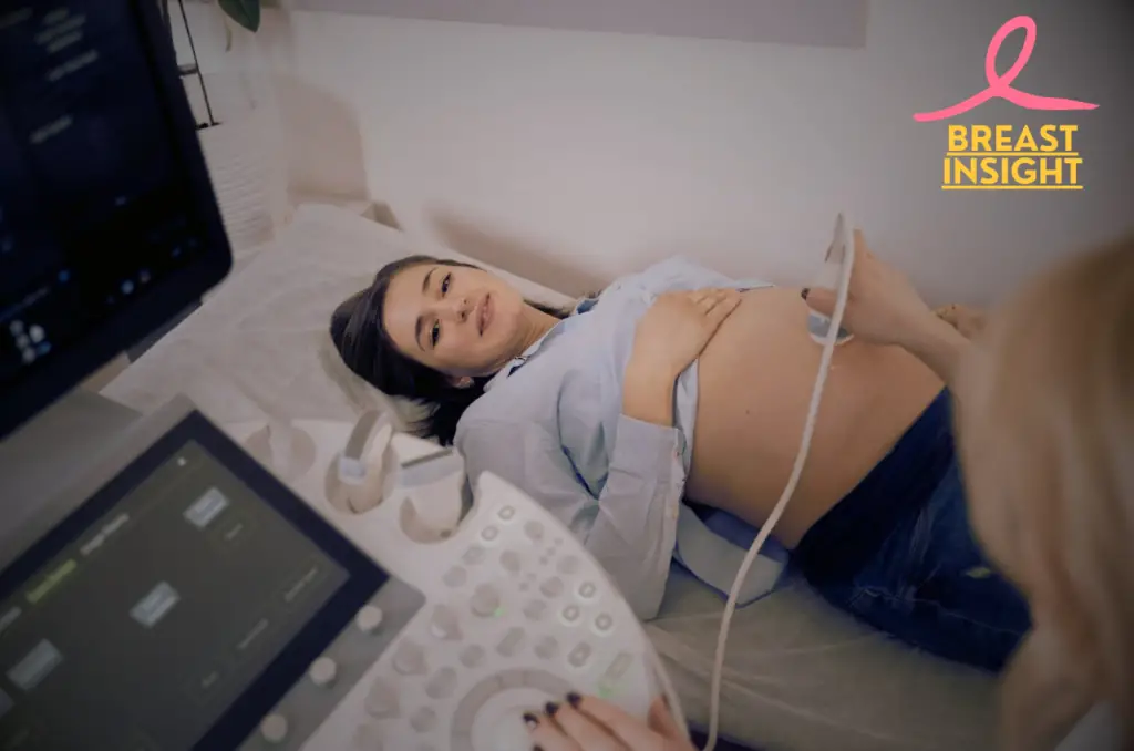
- The test usually lasts 15-30 minutes
- You will lie down on an examination table with your arm above your head
- A gel is applied across the skin to enhance the movement of sound waves
- A small gadget will be scanned over your chest and underarm area
- You might feel light pressure but no pain while undergoing the procedure
Preparation Guidelines
| Before the Procedure | Day of Examination |
| Wear comfortable clothing | Remove jewelry |
| Don’t apply lotions/powders | Wear a two-piece outfit |
| Bring previous imaging records | Arrive 15 minutes early |
| Continue medications as usual | Remove deodorant |
Results Interpretation
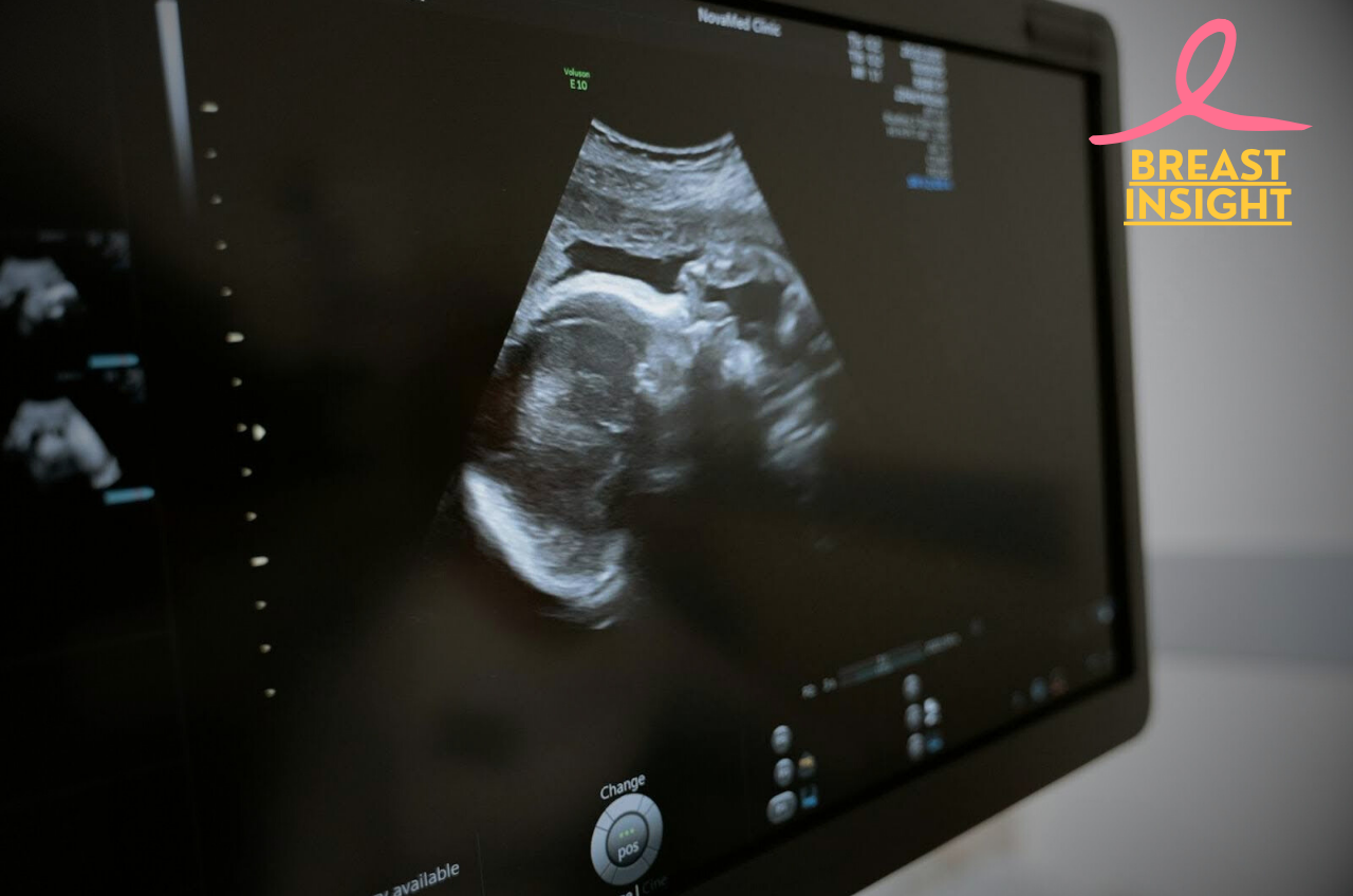
Real-time ultrasound images will be examined by a radiologist, who seeks
- Size and shape of problems with breasts
- Blood flow patterns
- Tissue composition changes
- Possible masses or cysts
Most results are reported using the BI-RADS scale, which ranges from 0 to 6. Your doctor will explain what your results mean and make any further recommendations for follow-up care.
If you feel a catch or pain, you can ask to be taken to see the sonographer. It’s an outpatient procedure, which means you can return to your other activities immediately. Your provider will make a follow-up appointment with you to discuss any results and options for your breast health.
Conclusion
Breast ultrasound is a very helpful tool in the fight against breast cancer. It is safe, does not involve cutting the skin, and does not use radiation for finding and checking on tumors. From simple ideas to more complex uses, ultrasound technology keeps improving. It gives doctors clear pictures that work well with mammography and make diagnosis more accurate. It can distinguish between a fluid-filled cyst, and a solid lump. Real-time images are also available, especially useful for younger women and even denser breast tissue.
With the vastly developing ultrasound imaging technology, the future would be easy for patients and help doctors to diagnose better. One may plan an appointment for a breast ultrasound or want to get more information about breast health. This technology is just one part of a complete procedure for finding and preventing breast cancer. Regular check-ups, talking openly with doctors, and being aware of your breast health are very important for finding problems early and getting good treatment results.
Frequently Asked Questions (FAQs)
What is a breast ultrasound, and how does it differ from a mammogram?
An ultrasound of the breast is a non-invasive, safe imaging technique that creates clear images with the aid of sound waves in the breast tissue. Unlike mammograms, which involve low levels of X-rays, ultrasounds do not use radiation and are therefore safer for some groups, primarily pregnant women. Ultrasound is often used in conjunction with mammography, especially in women whose breast tissue is very thick, or, more importantly, examine specific areas of interest during mammography.
Is a breast ultrasound painful?
Most patients state that the breast ultrasound is a painless test. Some may have minor feelings of pressure from the machine, but this is rare in terms of real pain. The gel put on your skin will feel cool, but the whole experience is far less invasive than other tests, such as biopsies.
How often should I get a breast ultrasound?
The frequency will depend on the individual risk factors and the guidelines of the healthcare provider. In addition to mammograms, some women with dense breast tissue or a personal or family history of breast cancer or other risk factors may benefit from annual breast ultrasounds. Therefore, this really must be determined in consultation with your healthcare provider about your best screening schedule.
What should I wear to a breast ultrasound appointment?
Patients are advised to wear comfortable clothing and avoid using deodorants, lotions, or powders on the day of the ultrasound, as these substances can interfere with imaging quality. It’s usually recommended to wear a two-piece outfit to make it easier to remove clothing from the waist up during the exam.

1 thought on “6 Eye-Opening Facts About Breast Ultrasound: From Basics to Breakthroughs”