In the United States, every two minutes, a person is diagnosed with breast cancer. However, when detected early through screening, the five-year survival rate for a breast cancer patient is 99%.
Despite these facts, many women are not willing to make an appointment to have their mammograms. It may either be fear of pain, lack of understanding about when to start screening, or confusion of different testing modalities. These fears keep most women from doing potentially lifesaving screenings. But one can imagine what relief it is to know the status of one’s being by getting checked regularly.
Understanding breast cancer is a crucial step toward overcoming these fears and taking proactive measures. If you’d like to learn more about what breast cancer is, its causes, symptoms, and available treatments, check out our in-depth guide here.
In this comprehensive report, we shall explore each and every relevant aspect of breast cancer screening as well as understand a few screening methods and look into advanced technologies enhancing the detection accuracy. Let’s break down the complexities involved in breast cancer screening and arm you with all the knowledge you need to make intelligent health care decisions.

Understanding Breast Cancer Screening Methods
Mammography and How It Works
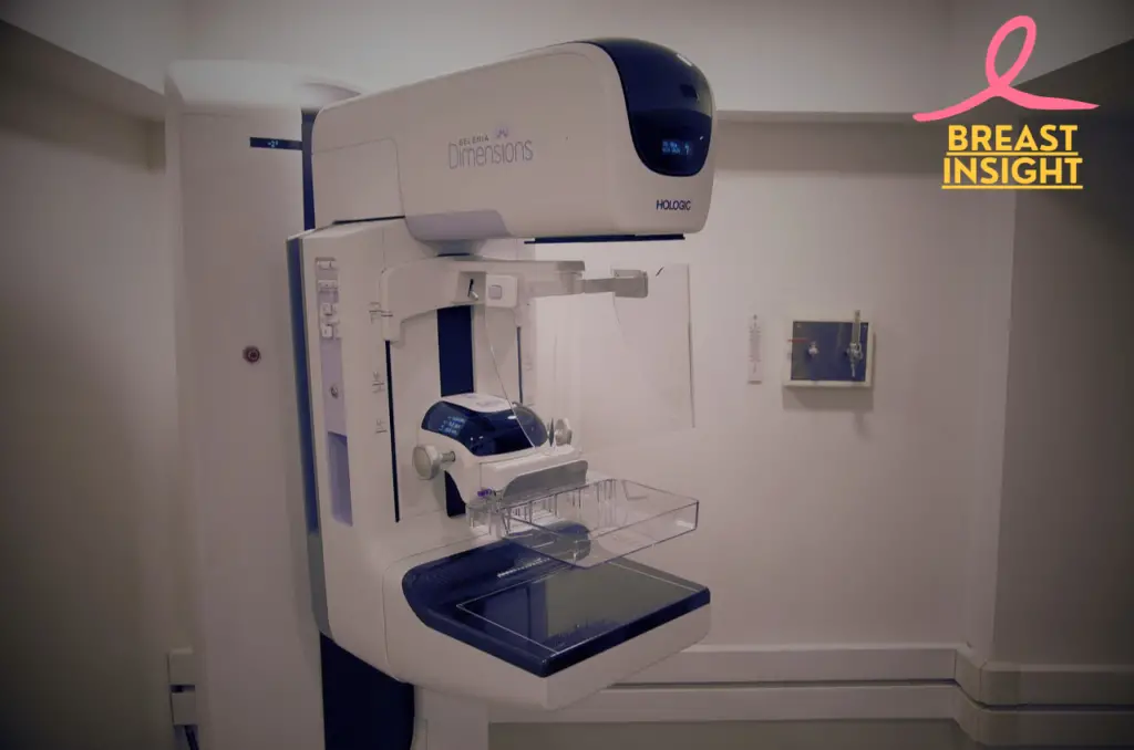
Mammography is the mainstay of breast cancer screening. Low-dose X-rays are used to generate detailed images of breast tissue. Modern mammography machines can capture 2D and 3D (tomosynthesis) images, which allows radiologists to preview the layer by layer of breast tissue.
| Type | Description | Benefits |
| 2D Mammogram | Traditional X-ray images from two angles | Standard screening tool, widely available |
| 3D Mammogram | Multiple X-ray images from various angles | Better for dense breast tissue, fewer false positives |
Clinical Breast Examinations
Clinical breast examinations (CBEs) are physical examinations conducted by healthcare professionals who systematically examine for
- Changes in breast size or shape
- Skin dimpling or abnormalities
- Nipple discharge or inversion
- Lumps or thickening in the breast tissue
- Lymph node changes in the armpit area
MRI Scan of High-Risk Individuals
Magnetic Resonance Imaging, or MRI, can create a comprehensive breast image by using magnetic fields and radio waves. It is useful for:
- Women with BRCA1 or BRCA2 genetic mutations
- Those with a significant family history of breast cancer
- Those who underwent chest radiation treatment prior to age 30
- Women having denser breast tissue
Ultrasound serves as a supplementary tool.
Breast ultrasound employs the use of sound waves to gather images of real-time structures and is an important adjunctive screening that contains the main uses;
| Application | Purpose |
| Dense Breast Evaluation | Distinguishing between normal tissue and suspicious areas |
| Targeted Investigation | Examining specific areas of concern found in mammograms |
| Guided Biopsies | Providing real-time imaging during biopsy procedures |
When used together, these screening methods form a holistic approach for the very early detection of breast cancer. Each mode has unique strengths and application, so they become precious tools in specific circumstances. Providers generally recommend a tailored approach to screening, taking into account the risk factors, age, and density of the breast tissue.
Breast cancer screening is only one aspect of the journey, as life after diagnosis can involve emotional and physical challenges. If you’re curious about navigating your sexual and emotional health after breast cancer, explore our helpful guide here.
Having understood the different screening methods, it is now time to consider when regular breast cancer screening should begin and how often such tests should be carried out.

When to Start Screening
Age-Based Recommendations
Current guidelines in medicine give age-specific recommendations for the screening of breast cancer. Here is a broad summary:
| Age Group | Screening Recommendation | Frequency |
| 40-44 | Optional, based on personal choice | Annual if chosen |
| 45-54 | Strongly recommended | Annual |
| 55+ | Recommended | Annual or biennial |
Women should consult their healthcare providers about starting mammograms at age 40; since evidence clearly demonstrates that early detection significantly improves treatment outcomes.
Risk Factor Measurement
Numerous factors determine the appropriate timing for initiating screening prior to standard recommendations:
Personal risk factors:
- Dense breast tissue
- Previous breast biopsies
- Previous radiation therapy to chest
- Hormone replacement therapy use
- Obesity
- Alcohol consumption
Healthcare service providers calculate individual risk scores with the help of risk assessment tools to guide an appropriate screening schedule.
Family History Considerations
Family history significantly impacts screening timing and frequency. Consider earlier screening if you have:
- First-degree relatives (mother, sister, daughter) diagnosed with breast cancer
- Many family members have either breast cancer or ovarian cancer
- Male relatives diagnosed with breast cancer
- Established genetic mutations in family (BRCA1/BRCA2)
Male breast cancer, although less common, also plays a critical role in family history. If you’re interested in understanding more about male breast cancer and its implications, check out our comprehensive guide here.
Recommendations for high-risk individuals:
| Risk Category | Screening Start Age | Additional Screening Methods |
| Strong family history | 5-10 years before earliest diagnosis in family | MRI + Mammogram |
| BRCA mutation carriers | Age 25-30 | MRI + Mammogram |
| Previous chest radiation | 8-10 years after radiation exposure | MRI + Mammogram |
Women who have such risk factors should undergo genetic counseling to establish an ideal schedule to use. Routine visits to healthcare professionals ensure that plans to undergo screening are tailored to individual risk profiles. Having the knowledge of when to start screening, let’s explore the numerous benefits that regular screening provides in maintaining breast health.

Advantages of Regular Screening
Early Diagnosis Benefits
- The earlier identification of suspicious masses
- Higher chances to detect the cancer before they spread
- Less invasive treatment modalities required
- Reduced likelihood of lymph node involvement
- An increased possibility for breast-conserving surgery
It can detect breast cancer from as early as 2-3 years from when actual physical symptoms would have occurred, thereby drastically improving the prognosis. Screening mammography has been proven to cut breast cancer mortality among women aged 40-74 by 20-40%.
Survival Rate Statistics:
| Stage at Detection | 5-Year Survival Rate |
| Stage 0 or I | 99% |
| Stage II | 85% |
| Stage III | 66% |
| Stage IV | 27% |
These statistics depict on their own the significance of early detection, through regular screening, regarding survival outcomes. For readers seeking more information about late-stage breast cancer, including its signs, treatment options, and survival rates, explore our in-depth guide here.
Treatment Options When Caught Early
Early stage diagnosis through timely screening offers more treatment options:
Less Invasive Surgical Alternatives:
- Lumpectomy Instead of Mastectomy
- Sentinel node biopsy versus complete axillary dissection
- Shorter recovery time
Reduced Need for Aggressive Treatments:
- A lower chance of requiring chemotherapy
- More targeted options of radiation therapy
- Higher aesthetic results
Cost-Efficiency of Prevention
Regular screening indeed proves to be cost-effective in the following ways:
- Reduced treatment expenses for early-stage cancer
- Reduced hospitalization time
- Fewer complications, requiring additional care
- Reduced time spent away from work during treatment
- Reduced need for extensive remodeling
Studies indicate that treating early-stage breast cancer costs approximately 30-50% less than treating advanced-stage disease. The average cost savings per case detected early ranges from $30,000 to $50,000.
Most health insurance plans include mammograms for screening under preventive care. Medicare and Medicaid also cover routine mammograms on a regular basis while fully cognizant of the process as important for public health. Having established the considerable benefits associated with routine screening it is pertinent at this point to address several common concerns that may impede women’s access to their recommended mammogram.

Addressing Common Concerns
Radiation Exposure Risks
Modern mammography equipment uses low amounts of radiation. All these are considered safe for routine screening purposes. Benefits of early detection far outweigh the minimal risks presented by radiation exposure. For perspective:
| Activity | Radiation Exposure |
| Digital Mammogram | 0.4 mSv |
| Natural Background (Annual) | 3.0 mSv |
| Chest X-ray | 0.1 mSv |
| Cross-country Flight | 0.04 mSv |
False positive results
While false positives can occur, they’re part of thorough screening protocol:
- About 10% of mammograms require additional imaging
- Most callbacks do not imply cancer
- Advanced Technology in the form of 3D Mammography Reduces False-Positive Reads by 40%
- Supplementary evaluations ensure accurate identification
Screening Discomfort
Most women experience minimal discomfort with mammograms:
- Compression lasts only a few seconds per image
- Scheduling during less sensitive times of month helps
- Experienced technologists can alleviate the pain
- Recent advancements in paddle designs enhance comfort without compromising image quality
Insurance Coverage
Breast cancer screening remains well-covered.
- The Affordable Care Act requires insurance to cover screening mammograms
- Medicare pays for annual mammograms for women 40+
- Numerous state programs provide complimentary screenings for women who lack insurance
- Local health departments typically offer some degree of low-income patient support
Time Required
New screening processes have become streamlined.
- Actual mammogram takes 15-20 minutes
- Most centers have flexible scheduling
- Results typically obtainable within 1-2 weeks
- Online portals provide direct access to the results
- Many centers offer same-day results
Screening is done as a norm whenever one understands the process and prepares for it. Additionally, incorporating self-awareness practices can enhance early detection efforts. Learn how to perform proper breast self-examination here. The temporary discomfort of screening is greatly surpassed by the benefits of early detection.
Up-to-date technologies in screening have improved the detection of breast cancer with improved accuracy and patient experience. Next, we delve into how these high-technology advancements are making screening for breast cancer more effective than ever.
Latest Screening Technologies
Advanced 3D Mammography
Three-dimensional mammography, commonly referred to as digital breast tomosynthesis, signifies a substantial advancement in the detection of breast cancer. This innovative technology generates multiple thin-layer images of breast tissue, thereby providing the following key advantages:
| Feature | Traditional Mammography | 3D Mammography |
| Image Detail | Single 2D image | Multiple layered images |
| Detection Rate | 4-5 cancers per 1000 | 6-8 cancers per 1000 |
| False Positives | Higher rate | 40% reduction |
| Tissue Overlap | Common issue | Minimal overlap |
Detection Using Artificial Intelligence
AI-powered screening systems are revolutionizing the detection of breast cancer through:
- Machine learning algorithms that analyze mammogram images
- Pattern recognition for sensitive anomalies
- Automated comparison with vast databases of past cases
- Real-time decision support for radiologists
The artificial intelligence implementation has shown outstanding results:
- 95% accuracy in initial screening
- 30% time saving in reading
- 50% reduction in unwanted callbacks
Mobile Screening Units
Mobile mammography units are bringing breast cancer screening into unprecedented accessibility.
Characteristics of Modern Mobile Units:
- Digital mammography machine
- Wireless data transfer
- Immediate image review capability
- Climate-controlled testing rooms
Benefits:
- Serves remote communities
- Reduces travel barriers
- Increases screening compliance
- Same-quality results as fixed facilities
These mobile units integrate cutting-edge technology while upholding high-quality standards. Outfitted with advanced digital mammography systems, many of these systems have 3D capability, which enables rural and underserved communities to receive care at the same level as that provided to patients in urban centers.
Recent research has shown that portable screening units increase screening by as much as 60% where it is not possible to access fixed facilities. It uses modern compression systems, which ensure that there are higher comfort levels during screenings without losing any qualities in the image.
If you’re navigating the challenges of metastatic breast cancer, explore our guide on Conquering Metastatic Breast Cancer: 5 Steps for actionable insights on living with and managing this condition.
Integration of the three technologies, namely 3D mammography, AI-assisted detection, and mobile units, has really enhanced the capture rates in breast cancer. All three are very important tools for early detection, which is still a most crucial factor for successful treatment. Indeed, these technologies are being developed further with frequent screening, promising much more accurate and accessible approaches for breast cancer detection in the future.

Conclusion
Regular screening still represents one of the best weapons in the war against breast cancer. From plain, two-dimensional mammograms to 3D imaging technologies, these methods help detect cancer early, when treatment is most effective. Understanding when to begin screening and maintaining a consistent schedule based on age and risk factors is crucial for your health.
Let common misconceptions about this health practice not stand in the way of pursuing it. Take a moment today to discuss a tailored screening plan with your health-care provider. Remember, early detection is life-saving-and controlling one’s breast health is one of the most important investments anyone could make towards their future.
Frequently Asked Questions (FAQs)
What age should women start getting breast cancer screenings?
Most health organizations suggest beginning annual mammograms at age 40. Women at increased risk due to a previous history of breast cancer or other causes, such as genetic predisposition-related causes-for example, BRCA mutations-will often want to start screenings earlier, typically at age 35. Regular discussions with a provider can help make the choice of appropriate screening schedule based on individual risks.
How often should I get a mammogram?
Women aged 40 to 44 years can start annual mammographies. From the age of 45 up to 54, a yearly check is recommended, and after the age of 55 years, biennial screening can be adopted or continued annually depending on someone’s preference. Again, those who have several risk factors may require more frequent screening. Always seek advice from a health provider.
Are there any risks associated with breast cancer screening?
A small risk with doing a mammography is breast cancer screening results in false positives and false negatives. False positives can leave patients under the torture of physical examinations and other diagnosis tests. False negatives essentially delay finding cancer. All the same, screening benefits in terms of early detection usually outweigh potential risks, making screening particularly important for women’s health.
What should I do if my mammogram results are abnormal?
If your mammogram results indicate abnormal findings, your healthcare provider will likely recommend additional tests, such as a follow-up mammogram, ultrasound, or biopsy, to determine the nature of the findings. It’s important to follow up promptly and discuss any concerns with your doctor to ensure a clear understanding of the next steps.
